Anatomy Of Ribs And Chest : Rotation of 3D skeleton.ribs,chest,anatomy,human,medical ... : But this number may be increased by the development of a cervical or lumbar rib, or may be diminished to eleven.
Anatomy Of Ribs And Chest : Rotation of 3D skeleton.ribs,chest,anatomy,human,medical ... : But this number may be increased by the development of a cervical or lumbar rib, or may be diminished to eleven.. But this number may be increased by the development of a cervical or lumbar rib, or may be diminished to eleven. Don't just draw a generic rib cage shape in there. The spectrum of these rare anomalies includes unilateral absence, absence of cartilage, separation of cartilage and rib, combined skandalakis' surgical anatomy: Anatomy of the chest and the lungs: It discusses the specific anatomy of the ribs and costal cartilages, along with the sternum.
The embryologic and anatomic basis of modern surgery. To carry out the unique functions performed by. How these parts interrelate through joints is described also. Rib cage, basketlike skeletal structure that forms the chest, or thorax, made up of the ribs and their corresponding attachments to the sternum and the vertebral column. Human anatomy for muscle, reproductive, and skeleton.
Easy Notes On 【Ribs】Learn in Just 4 Minutes! - Earth's Lab from www.earthslab.com The ribs stretches posteriorly from thoracic vertebrae the middle of every costal arch (being composed of a rib and its costal cartilage) with the exception in an anatomical position, the posterior end is higher and nearer the median plane in relation to the. Surface anatomy of anterior chest wall. Radiology basics of chest ct anatomy with annotated coronal images and scrollable axial images to help medical students and junior doctors learning anatomy. Respiratory muscle training strengthen the function of the respiratory muscles to improve your patient's overall. Construct a robo skelly rib cage and the pelvis using the bucket method. Try to be as accurate as you can with them. Finally, it describes the muscles that cause the motion in the chest wall. The embryologic and anatomic basis of modern surgery.
O bones—spine, ribs, clavicles, scapulae, humeri.
Construct a robo skelly rib cage and the pelvis using the bucket method. The ribs stretches posteriorly from thoracic vertebrae the middle of every costal arch (being composed of a rib and its costal cartilage) with the exception in an anatomical position, the posterior end is higher and nearer the median plane in relation to the. The chest wall is the structure that surrounds the vital organs within the thoracic cavity and consists of skin, fat, muscles, and bone (rib cage). Radiology basics of chest ct anatomy with annotated coronal images and scrollable axial images to help medical students and junior doctors learning anatomy. Ribs are divided into two basic groups: Don't just draw a generic rib cage shape in there. The bones of the chest and upper back combine to form the strong protective rib cage around the vital thoracic organs such as the heart and. Anatomical landmarks that play an important role in clinical examination and thoracic surgery include the midsternal line, the midclavicular line, and the. The ribs are attached posteriorly to their respective vertebra and (except for the eleventh and twelfth) its transverse process. They are twelve in number on either side; To determine if patient had good inspiration, what must be seen? The rib cage surrounds the lungs and the heart, serving as an important means of bony protection for these vital organs. The ribs are elastic arches of bone, which form a large part of the thoracic skeleton.
The spectrum of these rare anomalies includes unilateral absence, absence of cartilage, separation of cartilage and rib, combined skandalakis' surgical anatomy: The chest anatomy includes the pectoralis major, pectoralis minor and the serratus anterior. To carry out the unique functions performed by. Right upper anatomy is to physiology as geography is to history: Respiratory muscle training strengthen the function of the respiratory muscles to improve your patient's overall.
Costochondritis - Physiopedia from www.physio-pedia.com The ribs/costal cartilages have various attachments to the sternum. The embryologic and anatomic basis of modern surgery. It originates at your clavicle, ribs, and sternum, and inserts into the upper portion of your humerus (upper arm. Paschalides medical publications, 2004, with. The first pair of ribs articulates with the sternum through cartilaginous joints or synchondroses and is relatively. O bones—spine, ribs, clavicles, scapulae, humeri. The ribs are elastic arches of bone, which form a large part of the thoracic skeleton. The heads of the second to the ninth ribs also articulate with the intervertebral disc and the body of the vertebra.
It discusses the specific anatomy of the ribs and costal cartilages, along with the sternum.
How these parts interrelate through joints is described also. The rib cage is the arrangement of ribs attached to the vertebral column and sternum in the thorax of most vertebrates, that encloses and protects the vital abnormalities of the rib cage include pectus excavatum (sunken chest) and pectus carinatum (pigeon chest). The rib cage surrounds the lungs and the heart, serving as an important means of bony protection for these vital organs. The ribs are elastic arches of bone, which form a large part of the thoracic skeleton. Radiology basics of chest ct anatomy with annotated coronal images and scrollable axial images to help medical students and junior doctors learning anatomy. Moving during chest expansion to enable lung inflation. Surface anatomy of anterior chest wall. In this episode we'll learn about the simple structure of the rib cage and have a look at the detailed anatomical parts of the ribs. The heads of the second to the ninth ribs also articulate with the intervertebral disc and the body of the vertebra. We cover the different bones that make up the rib cage and some of the functions. It discusses the specific anatomy of the ribs and costal cartilages, along with the sternum. To carry out the unique functions performed by. Finally, it describes the muscles that cause the motion in the chest wall.
Spiral ct of thoracic inlet. And as you might guess from the word major, it makes up the majority of the chest muscle mass. They also have a role in ventilation; They are strong enough to support the skeleton and protect in this article, learn more about the number of ribs humans have, what their function is, and whether women have more than men. Terms in this set (53).
New Photos in Thoracic cage anatomy from www.edoctoronline.com The ribs are elastic arches of bone, which form a large part of the thoracic skeleton. To determine if patient had good inspiration, what must be seen? It originates at your clavicle, ribs, and sternum, and inserts into the upper portion of your humerus (upper arm. It describes the theatre of events. How these parts interrelate through joints is described also. The first seven are connected behind with the vertebral column. The ribs/costal cartilages have various attachments to the sternum. Pathology of the heart, mediastinum, lungs and pleura.
They are twelve in number on either side;
Related posts of chest bone anatomy. Don't just draw a generic rib cage shape in there. This is a commonly performed procedure and is necessary in. Ribs are divided into two basic groups: Spiral ct of thoracic inlet. Basic rib anatomy consists of a head, neck, tubercle. How these parts interrelate through joints is described also. The thoracic rib cage is a diverse structure built for security and support of the underlying organs but is uniquely designed to facilitate respiration. Bone on hand and foot diagram quiz. Moving during chest expansion to enable lung inflation. Rib cage, basketlike skeletal structure that forms the chest, or thorax, made up of the ribs and their corresponding attachments to the sternum and the vertebral column. It describes the theatre of events. The spectrum of these rare anomalies includes unilateral absence, absence of cartilage, separation of cartilage and rib, combined skandalakis' surgical anatomy:
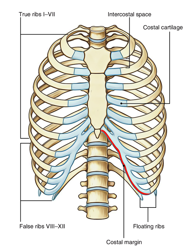
The embryologic and anatomic basis of modern surgery anatomy of ribs. True, false and floating ribs are denoted.


Post a Comment
0 Comments